Saturday, July 30, 2016

Remote control of tissue tolerance by regulatory T cells

This week journal PNAS published an interesting study on Foxp3+ regulatory T cells (Tregs). It showed that presence of Tregs within secondary lymphoid organs (in lymph nodes) was sufficient to prevent peripheral tissue immunopathology.

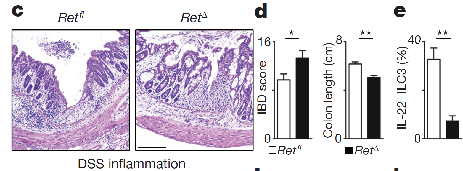
As a starting point for this study, the authors showed that mice with Tregs-specific deficiency of kruppel-like factor 2 (KLF2) develop non-fatal peripheral tissue immunopathology, even though in vitro such Tregs [Foxp3-cre; Klf2fl/fl] displayed normal suppressive activity).


Interestingly, in vivo adoptive transfer model, KLF2-KO Tregs also failed to prevent colitis and weight loss when co-transferred with WT naive T cells [but not with KLF2-KO naive T cells].


These results suggested that Tregs re-circulation between tissue and lymphoid tissues could have been involved. Surprisingly, the authors found no difference for Tregs presence [%-wise, no data about #] between KLF2-KO mice or WT (this is probably why this article ended up in PNAS).


Finally, the authors showed that combined deficiency of KLF2 and CCR7 in Tregs accelerated tissue immunopathology [by preventing Tregs access to lymph nodes].


In summary, the authors speculated that Tregs access to lymphoid tissue played a crucial role in preventing tissue immunopathology.

David Usharauli

Wednesday, July 27, 2016

Brain generates an autonomous immune protection against infection

Vaccines save lives. However even for most of vaccines that work we don't know why they work, i.e. we have no clue about protection correlates. They just work. Only source of information in human vaccine studies that are readily available is antibody titre analysis in serum [in blood]. But more recent studies in mice question the whole premise of vaccination strategy and analysis of its [correlates of] effectiveness. 

I am talking about tissue resident memory T cells. Several studies now clearly showed that most of the protection against infection is driven by local tissue-resident memory T cells rather than memory T cells found in circulation. For example, new study in Journal of Experimental Medicine showed that protection against brain viral infection is due to local brain tissue resident memory CD8 T cells, CD8 TRM cells.

To test the role of brain CD8 TRM cells in protection against viral infection the authors used recombinant, attenuated strain of LCMV virus (rLCMV). Initially they showed that unlike i.v. injection, when rLCMV  strain was administered directly in the brain (intrathecally) it led to robust development brain CD8 TRM cells.  


Moreover, when these mice were later challenged with WT virulent LCMV strain, mice with brain CD8 TRM cells showed superior control of virus (development of protective function of brain CD8 TRM cells were Ag-specific, IFN-γ and perforin-dependent, but CD4 and NK cell independent).


To further test the role of brain CD8 TRM cells in viral protection, the authors depleted circulating CD8 T cells with antibody. Still, these mice were fully protected against viral challenge with just brain CD8 TRM cells (Ab depletion does not affect tissue resident T cells). These data suggested that brain CD8 TRM cells were providing superior and independent protection against viral infection (though I don't think the authors have tested CD8 T cell depletion on mice injected with rLCMV intrathecally alone, because in most experiments on mice were done either with i.v. or i.v.+ i.th injections).


In summary, This study supports idea that correlates of some of viral [vaccine] protections could be found in tissues rather than in the blood.

David Usharauli

Tuesday, July 26, 2016

Infection-induced cell apoptosis activates self-epitope reactive T cells but barely

This week journal Nature Immunology published a study that had great title but poor data and conclusions. In fact, I rarely read such weak study for a long time, especially from top subject-matter journal. 

Basically, the authors tried to show that infection-induced cell apoptosis leads to self-peptide presentation and self-reactivity or even autoimmunity. Data are however misleading.

For this study the authors used the rodent pathogen Citrobacter rodentium that infects intestinal epithelial cells and induces their apoptosis. As a control, they have used infection with ΔEspF Citrobacter rodentium, a variant that lacks the secreted protein EPEC that mediates apoptosis. Initially, they showed that infection with WT Citrobacter rodentium, but not ΔEspF Citrobacter rodentium, induces Th17 response from large intestinal lamina propria (LI LP).


Next, the authors tried to examine whether infection-induced apoptotic cells will also provide self peptides for T cell activation (alongside of Citrobacter rodentium peptides). To do it, they have used so called double transgenic (DTg) mice derived from crossing OT-II mice with Act-mOVA mice. Now, these DTg mice delete absolute majority of OVA-specific OT-II cells in the thymus (from 1.5x10^6 to ~1,000 cells, i.e >1000X fold reduction of auto-reactive cells). The authors noted that DTg mice did not spontaneously develop autoimmunity and were healthy.


Next, when DTg mice were infected with Citrobacter rodentium, some portion of those OT-II cells left in DTg mice responded to it by up-regulating IL-17. The authors did not quantify the number of responding self-reactive OT-II cells and dot plot analysis reveals that their numbers seemed extremely low (on contour plot analysis). Moreover, it is not even clear whether self-reactive OT-II were responding to self-antigen or simply to inflammatory cytokine milieu [homeostatically] since even un-infected DTg mice showed proliferation and IL-17 expression in LI LP self-reactive OT-II cells.


The authors also showed that when infected with Citrobacter rodentium DTg mice showed little spike in anti-OVA IgA response driven by OT-II cells. However, it is not clear whether this anti-OVA IgA response has any pathogenic role.


Still, the authors believed that Th17 OT-II cells generated in DTg mice upon Citrobacter rodentium infection played pathogenic role in gut inflammation. As a "proof" they provided H&E staining of sections of large intestine from wild-type and DTg mice on day 40 after infection. Now, if scale bar on this H&E staining is 250 μm on both sections, then it is obvious DTg mice intestine is almost 2x more swollen or inflamed. But the authors noted that "DTg mice did not exhibit altered susceptibility to C. rodentium relative to that of wild-type or OT-II mice" and OT-II depletion did not significantly modify gut inflammation. So it is not clear from these data whether anti-OVA IgA or Th17 response after Citrobacter rodentium infection were in fact driving those observed pathogenic changes in the DTg mice guts (use of IL-17KO OT-II cells would have provided some guidance on this matter).


In summary, in my view this study only showed that WT Citrobacter rodentium infection induces little Th17 response from self-reactive T cells, however it failed to show that such Th17 response had any consequential effect.

David Usharauli  

Sunday, July 24, 2016

Fasting or fasting-mimicking diet synergize with anti-tumor chemotherapy

Two new papers in Cancer Cell caught my attention this week. In these studies scientists showed that fasting or fasting-mimicking diet (low calorie diet or calorie restriction mimetics) synergize with anti-cancer chemotherapy in a T cell-dependent manner.

For example, in one study it was found that in mice 48h fasting (0 calorie, very harsh "diet") or application of calorie restriction mimetics such as hydroxycitrate (that reduces cellular protein acetylation, thereby increasing autophagic flux) synergized with such anti-cancer chemotherapies as mitoxantrone (MTX) in a CD8 T cell-dependent manner.


Interestingly, this calorie restriction mimetic (HC) could not further enhance anti-cancer effect of chemotherapy in regulatory T cell-depleted [anti-FR4 treated] mice (i.e. it worked by restraining Tregs function).


Another paper also showed that fasting-mimicking diet could also reduce toxic effect of chemotherapy on healthy tissue in WT or T cell-deficient mice (independent of its anti-cancer synergy).


In summary, these two papers support idea that controlled calorie restriction could provide benefits to cancer patients during chemotherapy.

David Usharauli


Saturday, July 23, 2016

Group 3 innate lymphoid cells express RET and respond to glia-neuron network

Few days back journal Nature reported very interesting study from Portugal. In there, the authors showed that intestine glial cells (which are neuron-satellites expressing the glial fibrillary acidic protein (GFAP) signal local innate ILC3 cells via tyrosine kinase receptor RET.

First, they showed that ILC3 express RET that receives signals from glial-derived neurotrophic factor family ligands.


Second, mice lacking RET specifically in ILC3 cells, Rorgt-CreRet fl/fl (RetΔ), displayed marked reduction of IL-22, a cytokine involved in epithelial homeostasis.


Third, RetΔ mice showed heightened T cell-independent susceptibility to chemical [DSS]-induced colitis and to intestinal infection with the attaching and effacing bacteria Citrobacter rodentium.


Fourth, it showed that glial cells (RFP, red) are located in close proximity to ILC3 cells (GFP, green).


Fifth, mice lacking MyD88 specifically in glial cells, Gfap-CreMyd88Δ mice, also displayed heightened susceptibility to DSS colitis, indicating cross-talk between TLR signaling, glial-derived RET ligands and IL-22 produced by ILC3.


In summary, this study revealed existence of a complex network of neuro-glial-immune interactions that result in optimal defense against intestinal irritation. 

David Usharauli


Immune system of APS1/APECED patients maintains "tolerance" by targeting disease-modifying self-cytokines

AIRE deficiency defines APS1/APECED syndrome for which autoimmune polyendocrinopathy and chronic mucocutaneous candidiasis are signature phenotypes. 


For this study, the authors (associated with biotech company ImmunoQure) screened serum samples from APS1/APECED patients, their healthy relatives or unrelated healthy volunteers. Using a ProtoArray displaying ~ 9000 recombinant human proteins or protein fragments, the  researchers found that, among other things, sera from APS1/APECED patients showed significant reactivity to IFN, IL-17 and IL-10 family cytokines.


These hypermutated anti-cytokine antibodies could efficiently neutralize corresponding cytokines both in vitro in a cell reporter assay and in vivo in an ear inflammation assay.


Remarkably, among APS1/APECED patients, those with high titres of anti-cytokine neutralizing antibodies did not progress to type I diabetes (T1D) even though both population groups harbored anti-islet auto-antibodies (to GAD65).


In summary, this study indicates that immune system of APS1/APECED patients could maintain "tolerance" to some degree by targeting disease-modifying "self" cytokines.    

David Usharauli


Thursday, July 21, 2016

PD-1 signaling assists regulatory T cells when Foxp3 is down

Foxp3+ regulatory T cells (Tregs) maintain peripheral tolerance to self. Several molecules expressed by Tregs, such as CTLA-4, play crucial role in maintaining this state of tolerance to self. PD-1 is another such molecule, though its role in Tregs function is less clear.


This study arose from unexpected observation in new gene-modified mice where GFP was inserted in Foxp3 locus (mice carrying the IRES-GFPcre reporter KI at the 3′ untranslated region of the FoxP3 gene, FoxP3-GFPcreKI). When these mice were crossed with PD-1KO mice, it was found that male offspring of such cross showed early death, reminiscence of Foxp3KO mice. This was surprising since PD-1KO mice ordinarily do not show such phenotype.


Further experimentation found that GFP insertion affected Foxp3 stability thus resulted in reduced Foxp3 levels in FoxP3-GFPcreKI mice.


Indeed, FoxP3-GFPcreKI/PD-1KO male mice could be rescued with transfer of WT Tregs irrespective of their PD-1 expression indicating that PD-1 function were dispensable Tregs with normal level of Foxp3.


Finally, the authors showed that absence of PD-1 could further destabilize "Foxp3-low" Tregs function (conversion into ex-Foxp3 Tregs) resulting in lethal autoimmunity.


In summary, this study revealed that PD-1 could contribute to Tregs function in situations that affects Foxp3 stability.

David Usharauli


Tuesday, July 19, 2016

Oncolytic virus expressing PGE2 inactivating enzyme display enhanced anti-cancer effect

Oncolytic virus therapy is a new method of cancer therapy. Its goal is to selectively target cancer cells sparing healthy cells. However, not many tumors respond to it and so far it has limited application.


Initially, the authors tested in vivo susceptibility of several cancer cell lines to oncolytic vaccinia strain (WR.TK-Luc+). Some  cancers were less susceptible (Renca, 4T1), some more (LLC, MC38).


Susceptibility to WR.TK-Luctherapy was T cell mediated as CD8 T cell depletion could abolish it.


To overcome inhibitory micro-environment within resistant tumors oncolytic virus (OV) expressing prostaglandin-inactivating enzyme was designed. Indeed, modified OV therapy improved cancer protection.


Moreover, when combined with anti-PD1 therapy, modified OV could protect mice from resistant tumor such as Renca tumor, even when applied on established tumor (aspirin had no effect at this stage).


In summary, this study confirms that sustained inactivation of local prostaglandine source could drastically improve anti-cancer effect of OV, especially when combined with checkpoint inhibitors.

David Usharauli


Anti-CD40 agonistic antibodies with enhanced FcgRIIB binding mediate superior tumor protection

CD40 molecule was an immunology superstar of 90s. Scientists thought they found a mechanism that controlled robust CD8 T cell and antibody responses. However, 20 years later, nothing much came out of those studies. In fact, scientists are still figuring out what is the best way to activate human CD40 molecule. 

For example, in new study published in Cancer Cell, Jeffrey Ravetch's group showed that in mouse model expressing transgenic human CD40, effectiveness of anti-hCD40 antibody correlated with enhanced binding to inhibitory FcγRIIB.  

The authors developed several variants of anti-hCD40 (clone CP-870,893, a fully human IgG2, one of the most potent agonistic CD40 mAb) with higher affinity and selectivity to human FcγRIIB (and minimal change in binding to FcγRIIA). Protein immunization confirmed that improved affinity to FcγRIIB correlated with effectiveness of CD8 T cell priming.


Moreover, enhanced affinity of anti-hCD40 antibody to FcγRIIB (V11 clone, for example) correlated with improved anti-cancer effect of anti-hCD40 agonistic antibody (in MC38 and B16 cancer models).


In summary, this study indicates that in mouse model of hCD40 / hFcγRIIB expression selective binding to hFcγRIIB is important for enhanced anti-cancer effect of anti-hCD40 antibodies.

David Usharauli

Thursday, July 14, 2016

Hybrid NKB cells are source of early IL-18


By screening for NK1.1+ cell population in different tissues, the authors noticed that spleen and mLNs [but not other tissues] contained cell population expressing CD19 and IgM, a canonical B cell markers.


Image flow analysis confirmed that both NK1.1 and CD19 markers were expressed by the same cells.


This new NKB cell population were absent in RAG-KO, IL-2RgcKO, B cell or NK-deficient mice, suggesting unique lineage (its precursors appear to express CD122, IL-2Rbeta chain).


In vitro functional analysis showed that NKB cells were innate producers of IL-18 and IL-12 (IL-12p40, most likely). Of note, NKB cells did not secrete antibody after stimulation.


In an in vivo infection model NKB cell-deficient mice (created by reconstituting lethally irradiated mice with Id2 and mMT deficient bone marrow cells) showed high susceptibility to L.M. infection.


Finally, the authors showed that IL-18 produced by wt NKB cells played important role in disease resistance against L.M. infection.


In summary, this study identified new type of innate cells expressing NK and B cell markers, called NKB cells, that were involved in early IL-18 production that contributed early waves of IFN-gamma from NK and ILC1 cells and resistance to L.M. infection. It is not clear what roles surface CD19 or IgM play in NKB cell functionality.

David Usharauli


Wednesday, July 13, 2016

ILC2 sustain antigen-independent allergic responses

Most allergies represent exaggerated type II immune responses driven by adaptive Th2 cells. At least, this is what we used to believe it. However, discovery of rare innate cells, referred as innate lymphoid cells (ILCs), is slowly changing our understanding of cellular responses underlying allergies.

It is clear now that in laboratory mice model of allergy, type 2 ILCs (ILC2) contribute significantly and non-specifically [it seems] in sustaining allergy to irritant-allergens.


When analyzed ILC2 response to IL-33 or papain in lung tissue, the authors found that ILC2 displayed a typical adaptive-like behavior (expansion, contraction, quiescence).


Importantly, when IL-33 primed mice were challenged with allergen one month later ILC2 showed heighten type II response to allergen (papain) but not to control (saline). This response was "allergen"[protease]-specific but antigen-independent. It is possible that primed ILC2 were responding to IL-33 [or IL-25] released during papain challenge.


Similar data were obtained from mice primed with fungal Aspergillus protease (ASP) allergen and challenged 3.5 months later with papain (but not to saline). Here too, primed ILC2 could be responding [indirectly] to IL-33 or IL-25 released by papain.


In summary, this study showed that at least in mice "primed" innate lymphoid cells retain "heightened" non-specific responsiveness to allergen "long-term" (up to 6 months). This could explain why adaptive TH2 cell targeting immunotherapies may not be fully successful because it ignores contributions from innate cells such as ILC2.

David Usharauli


Tuesday, July 5, 2016

Selective elimination of autoimmune B cells using novel CAR-T technology based on antigen decoy

Few days ago journal Science published very interesting study describing a novel approach of treating autoimmune [auto-antibody]-dependent diseases. Called chimeric auto-antibody receptor (CAAR) T cell technology, this method can selectively eliminate autoantigen specific B cells in autoimmune diseases such as Pemphigus vulgaris, lupus, Myasthenia Gravis, Graves diseases and so on.

This new technology is based on a simple idea: antigen decoy. For example, during pemphigus vulgaris autoimmune B cells secrete autoantibodies to the keratinocyte adhesion protein desmoglein (Dsg3) that causes severe skin inflammation. Short-term management of diseases is achieved by total B cell depletion, but disease returns. The authors reasoned that if engineered T cells would express Dsg3 as a CARs, such antigen decoy CAAR T cells would selectively engage disease-causing anti-Dsg3-specific auto-antibody producing B cells. Such interaction should eliminate only Dsg3 specific B cells, sparing normal, infectious-specific B cells.

Indeed, the authors showed that CAAR-T cells expressing Dsg3 as a CAR construct (EC1-4 CAAR) can selectively interact with Dsg3-specific autoimmune B cells in vitro.  


More importantly in vivo NSG mice experiments confirmed that Dsg3 CAAR T cells could selectively eliminate human Nalm6 CD19+ B cell line expressing autoimmune Dsg3-specific receptors (PVB28/F779). In addition the authors claim that Dsg3 CAAR-T cells did not interfere with normal human skin epithelial functioning expressing Dsg3's natural ligand desmocollins.


In summary, if the results of this study is confirmed by other groups it would open up a new path for treating auto-antibody dependent autoimmune diseases.

David Usharauli


Saturday, July 2, 2016

Improving CAR-T cell anti-tumor selectivity by targeting protein glycoforms

Development of CAR-T cells selectively targeting solid tumors have been challenging due to shared antigenicity between cancerous and healthy tissues. Even minute level of protein [if] expressed on healthy tissue could produce unacceptable side-effects as seen for example in trials with CAR-T cells targeting her2/neu.  

New study in Immunity suggested to target protein glycoforms (here using anti-Tn MUC1 CAR-T cells) instead to avoid cross-targeting of healthy tissue.

The authors speculate that cancer-specific glycosylation could produce cancer-specific glyco-proteins. They found that Tn glycoform of protein mucin 1 (Tn MUC1) is selectively expressed by human T cell leukemia and by many solid tumors and can be specifically detected by CAR-T cells incorporating variable heavy and light chains derived from 5E5 mAb (the University of Copenhagen has patented the 5E5 antibody and antigen epitope and the University of Chicago has filed a patent on the 5E5 CAR).


The authors first tested 5E5BBz CAR-T cells against human Jurkat leukemia cells in vivo in NSG immunodeficient mice and observed that it could double survival of tumor challenged mice.


More importantly, 5E5BBz CAR-T cells were also effective against solid tumor, pancreatic tumor cell line expressing Tn MUC1 glycoform (while the authors reported no side-effects in mice subjected to 5E5BBz CAR-T cells transfer, it is not clear whether human tissue in vivo would express it cryptically).


In summary, the authors believe that targeting glycosylation variants of protein "specifically" expressed on transformed cells could overcome cross-targeting of healthy tissue by CAR-T cells.

David Usharauli